
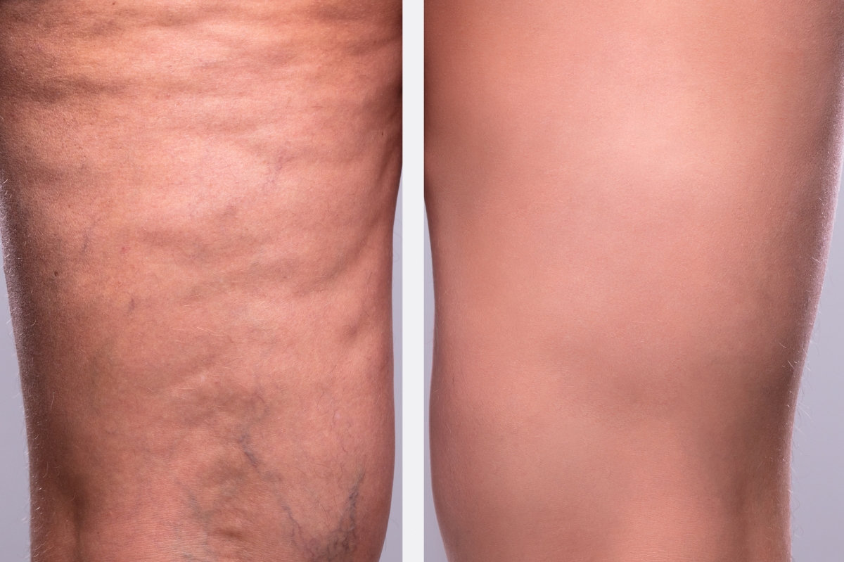
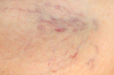
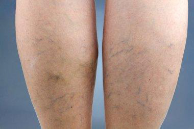
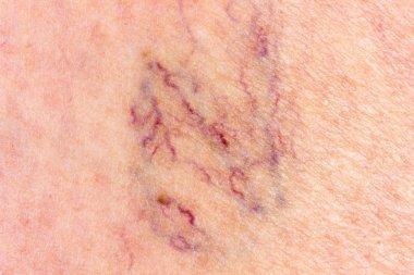
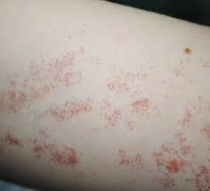
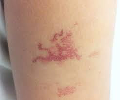
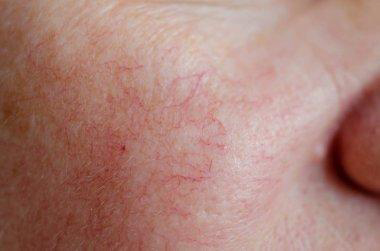
Spider veins (telangiectasias) are damaged, visible blood vessels just beneath your skin’s surface. They result from a weakening in the blood vessel wall or from faulty valves. They typically look red, blue or purple. They may appear in clusters that resemble spider webs or tree branches. Spider veins can form anywhere, but they usually develop in your legs or face.
There’s usually no need to worry. Spider veins aren’t dangerous by themselves. They simply mean some small blood vessels are damaged. Spider veins are mostly a cosmetic issue, meaning you might not like their appearance, but they won’t harm you.
Spider veins are common. They affect more than 50% of women and people assigned female at birth (AFAB), and they become even more common after age 80. Spider veins are more common among women and people AFAB compared with men and people assigned male at birth
Sometimes, though, spider veins are an early sign of chronic venous insufficiency (CVI). This is a vein disease that can affect your quality of life and lead to complications.
Telangiectasias need to be distinguished from other vascular conditions, including blood vessel tumors such as infantile hemangioma and angiomas that arise in adults; and capillary or venous vascular malformations.
Large red blood vessels are arteries and large blue blood vessels are veins. Arteries may be enlarged due to aneurysm formation. Veins enlarged due to the destruction of their valvular system are known as varicose veins.
Causes of spider veins:
In the legs, spider veins can occur when the valves inside the veins stop working correctly. Veins carry blood back to the heart. To prevent blood from flowing backward, they contain a one-way valve that closes once the blood passes through it.
If this valve weakens or sustains damage, the blood may have difficulty flowing in the correct direction, and it can begin to pool inside the vein. Over time, this can cause a bulge in the vein that branches out, resulting in spider veins.
Spider veins on the face are often the result of tiny blood vessels bursting. Increased pressure or sun damage can cause these to occur.
Factors that can increase a person’s risk of developing spider veins include:
- Genetics: Up to 90% of people with spider veins have a family history of them.
- Pregnancy: An increase in blood flow and the extra weight of the fetus on leg veins during pregnancy may cause spider veins.
- Sex: Spider veins occur in 41% of females ages 50 years and older. They tend to affect females almost twice as much as males.
- Age: The valves in veins tend to get weaker over time. The calf muscles, which help support the veins in the legs and enable them to pump blood upward, may also lose some of their strength as a person ages.
- Having overweight: Extra body weight can place added pressure on leg veins.
- Hormones: Hormonal birth control and hormonal treatments for menopause may increase the risk of spider veins. This is because estrogen can weaken vein valves.
- Sitting or standing for extended periods: Veins in the legs have to work harder to pump blood up toward the heart when a person remains in the same position for more than 4 hours
- Past blood clots or vein damage: This can damage the valves and make them unable to work correctly.
- Excess pressure in the face: This can be due to forceful coughing, sneezing, or vomiting. Some people may get spider veins on their face after pushing during childbirth.
- Sun damage: UV light from the sun can damage the skin and cause broken blood vessels or spider veins, especially on the face.
Certain medications may give rise to telangiectasia.
- Vasodilators especially calcium channel blockers; sun-exposed sites are mainly affected
- Long-term systemic corticosteroids
- Long-term topical corticosteroids, including steroid rosacea
- Intralesional triamcinolone injections
Telangiectasia may follow a cutaneous injury. For example:
- Scarring, including hypertrophic and keloid scars
- Livedoid vasculopathy and atrophy blanche
- Radiation damage
- Atrophie blanche
Classification:
Inherited conditions
Hereditary hemorrhagic telangiectasia: Hereditary hemorrhagic telangiectasia (HHT) is also known as Osler-Rendu-Weber syndrome. It is a rare inherited disorder that affects blood vessels throughout the body and is characterized by a tendency for bleeding in particular recurrent epistaxis (nosebleeds); and skin telangiectasia (skin lesions resulting from dilation of blood vessels).
The two major types of HHT are HHT1 and HHT2. They are caused by mutations in the endoglin (ENG) and activin receptor-like kinase type 1 (ACVLR1) genes found on chromosomes 9 and 12 respectively. Two other genes have also been identified. A defect in just one of these genes causes an abnormality in the formation of blood vessels, which may easily rupture and bleed. The abnormal blood vessels are known as Telangiectasias, or arteriovenous malformations (AVM) if larger blood vessels are involved.
The diagnostic criteria for HHT are:
- Spontaneous recurrent nosebleeds
- Multiple Telangiectasias on skin and mucous membranes
- Involvement of internal organs
- An affected parent, sibling or child
Benign hereditary telangiectasia: Benign hereditary telangiectasia is a primary telangiectatic disorder. It has varying clinical patterns. They are often widely distributed over the face, neck, upper trunk, dorsal, hands and, less frequently, knees. Telangiectasias may be macular, plaque-like, arborizing, reticulated, punctate, mottled, spiderlike, and linear.
- Punctate Telangiectasias may be surrounded by pale halos (steal phenomenon).
- Some lesions may be indistinguishable from spider telangiectasis.
- Early Telangiectasias can be small and erythematous. They often increase in size and may become paler with age.
- The mucosal membranes are not affected with the exception of the vermilion border of the lips.
- There is no systemic involvement or bleeding, in contrast to hereditary hemorrhagic telangiectasia.
- The Telangiectasias are invariably asymptomatic and cause only mild cosmetic concerns.
Generalized essential telangiectasia:
Generalized essential telangiectasia is just one type of primary telangiectasia and is given this name because of its widespread distribution pattern over the body. The cause of generalized essential telangiectasia remains unclear. Sometimes there may be a family history, which suggests a genetic component.
Unilateral nevoid telangiectasia: Unilateral nevoid telangiectasia is a rare congenital or acquired disorder. The majority of the authors propose that it is probably related to elevated estrogen receptors, although other authors advocate different theories.
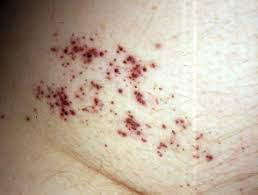
Angioma serpiginous: Angioma serpiginous is a skin condition in which there are small blood vessels near the skin surface. It presents as small red dots (puncta) that cluster together to form a linear or snake-like array (serpiginous pattern) or ring-shaped (gyrate) pattern. There is no bleeding, inflammation or pigmentation.
Rothmund Thompson syndrome: Rothmund–Thomson syndrome is a rare inherited disease that affects the skin, eyes, bones, and internal organs. At least 300 cases have been reported in medical journals since a case was first described by Rothmund in 1868. Rothmund–Thomson syndrome is also known as poikiloderma congenitale.
Rothmund–Thomson syndrome is due to a genetic defect, in which there are mutations in the RECQL4 gene on Chromosome 8. This gene encodes for the enzyme DNA helicase which unwinds DNA (deoxyribonucleic acid). The abnormal gene makes the chromosomes unstable, altering the growth of cells in many tissues.
Ataxia-telangiectasia:
Ataxia-telangiectasia is a rare inherited multisystem disorder that is characterized by:
- Ataxia (lack of co-ordination)
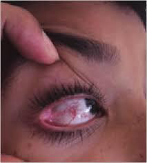
- Telangiectasias on the skin and eyes
- Severe combined immunodeficiency resulting in recurrent respiratory infections
- A predisposition to malignancy.
- Ataxia-telangiectasia is also known as Louis-Bar syndrome.
Ataxia-telangiectasia may occur in any race or sex. Reported rates of incidence range from 1 in 40,000 to 1 in 100,000 births. The incidence of ataxia-telangiectasia is significantly higher when parents are consanguineous, for example in the Bedouin population.
Bloom syndrome (congenital telangiectatic erythema): Bloom syndrome is an autosomal recessive inherited disorder, which means that two abnormal Bloom syndrome genes are needed for the disease to be apparent (one from each parent). If a person has one affected gene, he or she is called a carrier of Bloom Syndrome and does not manifest symptoms of the disease. If both parents are carriers, there is a 1 in 4 chance of having an affected child with each pregnancy. Recessive diseases are often consanguineous (eg, cousin marriages).
Bloom syndrome is more common in eastern European Ashkenazi Jews. At least 1 in 100 Ashkenazi Jews is a carrier of the disease. It appears to be slightly more common in males than females.
Cockayne syndrome:
Cockayne syndrome is a rare inherited disorder in which people are sensitive to sunlight, have short stature, and have the appearance of premature aging. In the classical form of Cockayne syndrome (Type I), the symptoms are progressive and typically become apparent after the age of 1 year.
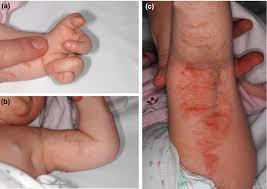
Cutis marmorata telangiectasia congenita:
Cutis marmorata is a condition where the skin has a pinkish blue mottled or marbled appearance when subjected to cold temperatures. Rewarming usually restores the skin to its normal appearance. Cutis marmorata occurs in about 50% of children and is typically seen throughout infancy. Adults may also be affected.
The mottled appearance of cutis marmorata is caused by superficial small blood vessels in the skin dilating and contracting at the same time. Dilation creates the red color of the skin whilst contraction produces a pale appearance. This phenomenon is most pronounced when the skin is cooled. The reasons for the reaction are not fully understood.
Focal dermal hypoplasia (Goltz syndrome):
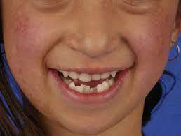
Focal dermal hypoplasia is a genetically inherited disorder that can affect the development of many different organ systems and was first described by Goltz in 1962. It presents with characteristic abnormalities of the skin, eyes and teeth and may also have effects on the skeletal, gastrointestinal, genitourinary, neurological and cardiovascular systems. The name is actually misleading, as thinning of tissues is not confined to the dermis but also involves the epidermis and subcutaneous tissue.
Kindler syndrome:
Kindler syndrome is a rare hereditary disorder, associated with skin fragility. The syndrome involves the skin and mucous membrane with radiological changes. The genetic defect has been identified on the short arm of chromosome 20.
Acquired telangiectasia
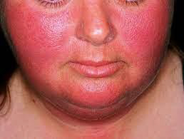
Rosacea: Rosacea is a chronic inflammatory skin condition predominantly affecting the central face and most often starts between the age of 30–60 years. Rosacea typically presents after the age of 30 and becomes more prevalent with age. However, it can occur at any age and occasionally presents in children. Although rosacea can affect anyone, it is more common in those with fair skin, blue eyes, and those of Celtic or North European descent. It may be more difficult and under-recognized in patients with skin of color.
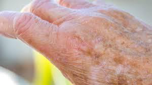
Sun damaged and aged skin especially in those who smoke:
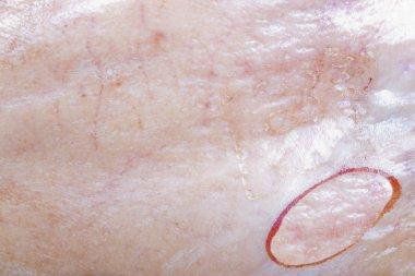
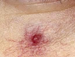
Spider telangiectasis (also called spider naevus): A spider telangiectasis is composed of dilated blood vessels, and is clinically characterized by its spider-like appearance. It is given that name because it has a central red papule (the body of the ‘spider’) from which fine red lines (the spider legs) extend radially. An alternative explanation for the name explains that the red lines form a spider-like network. Other names for spider telangiectasis are spider angioma (a misnomer), spider naevus and naevus araneus.
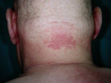
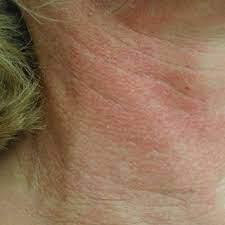
Poikiloderma of Civatte (sun damage affecting the sides of the neck): Poikiloderma of Civatte is a benign, common and chronic condition, which belongs to the group of melanodermas (pigmented skin disorders). The term ‘poikiloderma’ refers to a skin change with atrophy where hypopigmentation/hyperpigmentation changes and dilation of the fine blood vessels (telangiectasia) can be seen in the affected skin. Long-term sun exposure is considered to be a main contributing factor.
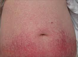
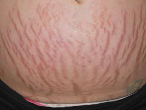
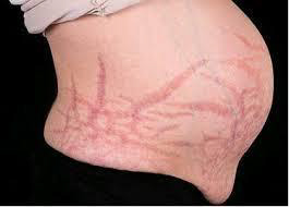
Pregnancy:
During pregnancy there are marked changes in sex hormones, the immune system, and the cardiovascular system, and this can lead to changes in the skin. Many changes in the skin, hair, and nails of pregnant women are common and considered normal (physiological).
Pigmentation in pregnancy
Linea nigra, darkening of pigmented areas (ie, nipples, areolae, genitals), and generalized increase in pigmentation appears in the first trimester of pregnancy. It affects 90% of pregnant women, particularly those with skin of color (Fitzpatrick skin types IV-VI). This pigmentation fades after delivery, but usually not completely.
Cushing syndrome: Cushing syndrome is a hormonal disorder caused by prolonged exposure to inappropriately high levels of plasma glucocorticoid (also referred to as cortisol) hormones. Glucocorticoid hormones maintain glucose regulation, suppress the immune response and are released as part of the body’s response to stress. The production of cortisol from within the cortex of the adrenal glands is regulated by a small gland just below the brain called the pituitary gland.
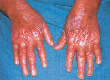
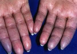
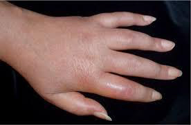
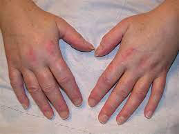
Systemic sclerosis especially CRST syndrome (which forms telangiectatic mats): Systemic sclerosis (SSc) is an autoimmune inflammatory condition. It results in potentially widespread fibrosis and vascular abnormalities, which can affect the skin, lungs, gastrointestinal tract, heart and kidneys. The skin becomes thickened and hard (sclerotic).
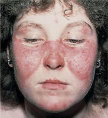
Cutaneous lupus erythematosus: Cutaneous lupus erythematosus (LE) is a diverse group of autoimmune connective tissue disorders localized to the skin that can be associated with systemic lupus erythematosus (SLE) to varying degrees.
Cutaneous lupus erythematosus has an annual incidence of 4 cases per 100,000 people, and a prevalence of 73 cases per 100,000.
As with SLE, there is a marked female predominance with CLE particularly affecting women 20 to 50 years of age. However, all age groups and both sexes can be affected. Skin of color is an important predisposing factor.
Mixed connective tissue disease: Mixed connective tissue disease is a rare autoimmune connective tissue disorder with anti-U1RNP auto-antibodies. The cause of mixed connective tissue disease is not fully known. Due to the presence of autoantibodies, it is classified is an autoimmune disease. It is most common in patients with HLA-DR4, suggesting it has a genetic background.
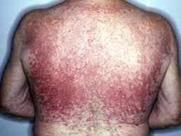
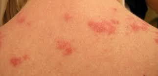
Dermatomyositis: Dermatomyositis is a rare disease that causes muscle weakness and skin rash. Symptoms include a red or purple rash on sun-exposed skin and eyelids, calcium deposits under the skin, muscle weakness, and trouble talking or swallowing. There is no cure, but treatment is done to reduce the symptoms.
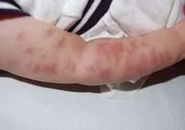
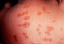
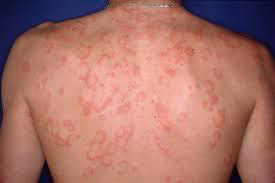
Mastocytosis, specifically the variant, Telangiectasia macularis eruptiva perstans: Mastocytosis is a diverse group of disorders characterized by the expansion and accumulation of mast cells in one or more organ systems. It can affect the skin, bone marrow, liver, spleen, gastrointestinal tract, or lymph nodes. In 2022, the World Health Organisation (WHO) categorized three main types of mastocytosis:
- Cutaneous mastocytosis (affecting the skin alone), most commonly maculopapular cutaneous mastocytosis
- Systemic mastocytosis
- Mast cell sarcomas
Mastocytosis can occur at any age, although some types are more common in particular age groups. Females have a slightly higher risk of developing mastocytosis than males. In most cases, there is no family history of mastocytosis.
Carcinoid syndrome:
Carcinoid syndrome is characterized by episodic cutaneous flushing, diarrhea, and wheezing. Carcinoid syndrome is the result of a combination of peptides and amines secreted by advanced neuroendocrine tumors (NETs) into the bloodstream. Carcinoid syndrome affects up to 19% of people with neuroendocrine tumors, which can be present in the gut, pancreas, lung, kidney, ovaries, and testes. They are rare in children. The incidence is 1–2 per 100,000 people annually.
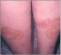
Necrobiosis lipoidica: Necrobiosis lipoidica is a rare granulomatous skin disorder typically described on the shin of diabetics. Necrobiosis lipoidica is three times more common in females than in males, and usually develops in young and middle-aged adults. Necrobiosis lipoidica begins as a dull red papule or plaque on the shin which slowly enlarges into one or more yellowish-brown patches with a red rim.
Male genital dysaesthesia:
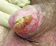
Male genital dysaesthesia is a form of cutaneous dysaesthesia characterized by a burning, hot, irritating discomfort of the penis, foreskin, and/or scrotum. It is often accompanied by increased sensitivity to touch.
Male genital dysaesthesia is also known as Dysaesthetic penoscrotodynia (DPSD) or male genital burning syndrome. If dysaesthesia is accompanied by redness, this may be referred to as red scrotum syndrome. Localized symptoms can be described as scrotodynia or burning scrotum syndrome.
Male genital dysaesthesia can affect males of all ages and skin color. It is more common amongst those with Fitzpatrick type 1 or 2 skin, and those over the age of 50. Genital dysaesthesia associated with redness and vascular hyperreactivity of the scrotum may be associated with rosacea.
Treatment
There are different treatment options a person can try:
- Compression stockings or socks
- Wearing compression stockings or socks places pressure on the veins in the lower legs. This pressure can help improve blood flow and prevent further spider or varicose veins.
- Compression stockings may also help relieve leg swelling and lower the risk of blood clots in the legs.
Types of compression stockings include:
- Support pantyhose: These provide only light pressure but are available in many stores.
- Gradient compression stockings and socks: These provide medium pressure around the feet, ankles, and calves. They are often available at specialist stores and pharmacies.
- Prescription compression stockings: These provide the most pressure to the feet and legs. They come in various sizes and strengths, as well as footless varieties. Prescription compression stockings are not suitable for some people, including those who have heart failure or other heart problems.
- Sclerotherapy and closure system: Sclerotherapy involves injecting an irritant directly into the affected vein. Due to irritation, the veins stick together and keep blood from flowing into the area. This procedure can reduce swelling and cause the vein to shrink. Over time, the spider vein fades or vanishes. Several treatments may be necessary to obtain the desired result.Similar to sclerotherapy, closure system treatment involves injecting a substance into the affected veins. This substance is sticky, and it closes the vein off from blood flow, causing the spider vein to fade or disappear with time.As with sclerotherapy, a person may require several treatments to produce the desired outcome.
- Laser treatment: A healthcare professional can use a laser to treat small spider veins close to the skin’s surface. The laser is a strong, focused beam of light that causes the spider vein to clot and dry up.Laser treatments are less invasive than sclerotherapy and closure systems because there is no injection. However, laser treatments are typically less effective in reducing spider veins.
- Endovenous laser therapy (EVLT): EVLT is a newer procedure for treating spider veins and small varicose veins. Endovenous means that the procedure is done inside the vein.A healthcare professional makes a small incision in the affected vein and then inserts a laser fiber. The laser applies heat directly to the vein and causes it to collapse. The vein may take several months or up to a year to disappear. EVLT involves the use of local anesthesia.
- Surgery: Although some surgical treatments can be effective for larger varicose veins, doctors typically do not perform them on spider veins.
Prevention
Certain lifestyle changes and self-care tips can help prevent new spider veins from appearing or stop existing ones from getting worse. These include:
- Wearing sunscreen: Applying sunscreen every day can help prevent spider veins, particularly on the face. Use sun-protective hats and clothing when outdoors for extended periods.
- Reaching or maintaining a moderate weight: This helps reduce pressure on the veins and keeps blood flowing well.
- Wearing compression stockings: If spider veins or varicose veins are a concern or run in the family, consider wearing compression stockings or socks.
- Staying mobile: Avoid sitting or standing for extended periods without taking a break.
- Avoiding tight clothing: Clothing that is too tight around the waist, legs, or pelvis can restrict blood flow and may increase the risk of spider veins.
- Avoiding overuse of hot tubs and saunas: Excessive heat can cause veins to swell, increasing the risk of dilated and bulging veins in the legs.
- Limiting alcohol consumption: Drinking alcohol can cause flushing in the face and broken blood vessels in some people.
- Getting regular exercise: Physical activity can help improve circulation and prevent blood from pooling in the legs.
- Elevating the legs: Raising the legs when sitting or lying down can help prevent blood from pooling downward in the legs.
- Contacting a dermatologist: People with skin conditions that can increase the risk of spider veins, such as rosacea, may want to consider contacting a doctor or dermatologist to discuss treatment options.
- Using cover-up products: If the appearance of spider veins is a concern, people may wish to use body or leg makeup to mask or minimize them temporarily. Self-tanning products can also work for this purpose.